What’s new? – September 2018

What’s new? – July 2018

My monthly overview of some of the papers from the literature I found interesting or important.

Your gut is talking, and your brain is listening

The fact that your digestive system communicates with your brain is nothing new. Your stomach tells your brain you’re full, your intestine might suggest it’s time to find a toilet soon. The gut has its own little brain, the enteric nervous system. Until recently, it was thought that the communication between the brain and the enteric nervous system was only indirect. The brain or the gut can secrete hormones and other signalling molecules, and these travel through the bloodstream to eventually have their effects.

Now, as reported in Science, Kaelberer et al show there is a far more direct route of communication. At least in the mouse, there is a direct nervous connection between the gut and the brain. The starting point was the observation that the signalling cells in the gut, the enteroendocrine cells, make peptides such as CCK and PYY which are normally released into the blood as hormones. However, these can also function as direct neurotransmitters at synapses.

Using a modified rabies virus, which only spreads through synaptic neuronal connections, they show these enteroendocrine cells do connect to a prominent nerve fibre called the vagus nerve, as well as the brainstem. The authors managed to recreate this system in a dish by culturing cells from the vagus nerve and this intestine, and not only showed that these wire up, but that the nerve cells respond to nutrients such as glucose. Through a series of experiments, they were able to confirm that it’s the enteroendocrine cells talking to the nerve cells causing this response.

Using optogenetics, a technique that allowed for cells to be turned on or off at will by exposing them to light, they show that activating or blocking the signalling from enteroendocrine cells modifies the firing rate of the vagus nerve. Doing this modifies the response to a glucose stimulus, and significantly affected the feeding behaviour of the mouse, showing this system has a real function. Far from just being an organ to absorb food, it seems more and more the case that the gut can have a real influence on our behaviour.


Microglia don’t like fat

Ok, I think we might be hitting peak microglia soon, they do seem to be involved in absolutely everything these days. This new study from Elisabeth Gould’s lab looks at the role of microglia in obesity. Something most people (including me!) might not have realised is that life-long obesity is associated with a decline in cognitive functioning, such as memory impairments.

In this study, mice were fed a high fat diet, leading to high levels of obesity (40 grams might not sound much, but it makes for a pretty chubby mouse….). These obese mice did perform worse on a number of cognitive tasks than the normal diet controls. Looking closely at the brains of these mice, they saw a decreased density of synaptic spines, or in other words, reduced numbers of connections between neurons.

As I’ve discussed before, one of the most prominent suggested non-immune functions of microglia is synaptic pruning, or the removal of weak neuronal connections. Could this process be going wrong here? Indeed, the authors saw increased activation of microglia in the obese animals, and more of them on or around synapses.

Genetically blocking microglial activation by knocking out the gene CXCR1 meant obese mice didn’t develop changes in neuronal connectivity or cognitive problems. Treating the mice with the anti-microglial drug minocyline, or inhibiting synaptic pruning with annexin-V for several weeks gave the same results, showing these deficits can be reversed.

So why would this be the case? It has been known for some time that chronic obesity can lead to low level inflammation. This can lead to higher levels of inflammatory proteins such as interleukin 6 and CRP. It is quite likely these inflammatory factors in the rest of the body turn on the microglia in the brain, which then in turn do neuronal damage. Would be a good hypothesis to test!


Hit the loop button to remember

As any student who has ever revised for an exam knows, repetition will help things stick. The brain knows this as well. There has been a lot of work in rodents where animals have been trained to run on a maze. This training is reflected by brain cells firing at specific places in a maze. When you record the activity of these cells when the animals sleep after the training, these exact same cells become active again, in the exact same order as if the animal was running on the maze again. If you disrupt these process, the animal won’t learn the maze as well. This phenomenon has also been seen in humans, brain areas that have been activated during a task also get activated in rest periods afterwards, and the hypothesis is that, as in rodents, this helps with consolidating memories.

In a cool study in Nature Communications, Shapiro et al present data that suggests this is in fact true. In the first session, volunteers were presented with a series of images of objects and asked to remember their specific features. Brain activity was measured by fMRI during the training and when subjects were tested on their memory of the objects. Afterwards, they were scanned while not seeing the subjects or being tested. Twelve hours later, this was repeated.

What they found was that interestingly, in the period after the testing, the brain activity patterns correlating to the objects that were remembered the worst were replayed by the brain the most. It is almost like the brain known which memories it needs more practice on. Evidently this practise works, as those memories which were replayed most often after session 1 were best remembered in the second session, 12 hours later.

However, this was only strongly the case in one situation; if the participants slept in between the sessions. If the two sessions were on the same day, there wasn’t anywhere near as strongly an effect of replay. So, all in all this would suggest there is a scientific reasoning for the old advise to keep revising and get a good night sleep before an exam.


Neurogenesis as a therapeutic mechanism? Hit the ground running

The formation of new neurons, or neurogenesis, in the adult brain is limited and restricted to certain areas such as the hippocampus – this is why losing neurons is such a big deal, most of the time, you can’t replace them. Hippocampal neurogenesis is required for a variety of memory processes and is disrupted in many diseases. There have been attempts to increase neurogenesis as a therapeutic strategy, mainly in depression, but so far this has not been successful.

Writing in Science earlier this month, Choi et al show an approach that might change this. Their study uses a transgenic mouse model of Alzheimer’s disease (how valid these models are is a good question, but that is a different debate). These animals start showing learning and memory problems as they age. The authors show that just increasing hippocampal neurogenesis with drugs either did not improve the performance of the animals in memory tasks, or showed at most a modest improvement, depending on the sex of the animals and the specific task.

However, if the treatment was combined with exercise, which is also known the stimulate neurogenesis, there was a much more robust increase in performance. On the other hand, exercise alone did not cause an improvement either, only the combination did. In the reverse experiment, they showed that blocking neurogenesis worsened task performance.

So why is this combination effective when the individual treatments aren’t? It is known that exercise increase the levels of BNDF, a factor which supports the survival of neurons. The authors showed that increasing BNDF levels mimicked the actions of exercise. Although they do not directly show it, the idea is that you require the beneficial effects of exercise, through increased BDNF levels to help the new neurons survive and give a functional improvement.

As good as that seems, the increases in test performance are still modest, and getting this to work in humans would be a challenge. Nevertheless, interesting data.


Stay tuned for more posts here on Neuroscience Ramblings, and in the mean time, follow me on Twitter: @DrNielsHaan

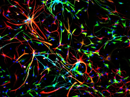
 

Fresh from the lab: our latest work on microglia and adult neurogenesis in schizophrenia

Our latest preprint has gone live on bioRxiv. This is some work we’re very excited about, so I thought I’d talk a bit about it here

You can find out latest preprint, “Haploinsufficiency of the schizophrenia risk gene Cyfip1 causes abnormal postnatal hippocampal neurogenesis through a novel microglia dependent mechanism” here. This work, which I did most, but certainly not all the work for, links three very different areas of research together, areas which have not been linked before. These are adult neurogenesis, genetic risk for schizophrenia (and other psychiatric diseases), and microglia.

Most of the brain can’t make any new cells in adulthood. However, the hippocampus is an exception to this rule. Here, in a structure called the subgranular zone of the dentate gyrus sit neural stem cells, which can make new neurons throughout life in a process called adult hippocampal neurogenesis. These adult born neurons are specifically used in several functions of the hippocampus, mostly to do with memory. Interestingly, there is some evidence hippocampal neurogenesis is disrupted is psychiatric patients, including in schizophrenia, and might be involved in some of the symptoms of these diseases.

Making new neurons in the adult brain broadly requires four phases. The first is cell division. This is when the stem cells that sit in the subgranular zone divide to generate new cells that will eventually become neurons. The second phase is differentiation. This is when a cells slowly change from a stem cell to something that will become a neuron. Call it neuronal puberty. In parallel with this, the third phase starts, which is cell migration. The developing neurons move from their birthplace into the adjacent granular zone to their eventual final location. This is where the last phase takes place, which is maturation. This is when the new neuron wires into the exiting circuitry and starts functioning. In rodents, this process takes up to eight weeks.

As a lab, we use a lot of genetic models of schizophrenia. Now, I’ve written about schizophrenia models before, so I’m not going to go on about their validity. What we used here was a mouse model for a human genetic risk factor called the 15q11.2(BP1-BP2) CNV. This means carriers have a small deletion in one copy of chromosome 15. Carrying this deletion doubles your risk for schizophrenia, as well as increasing risk for autism, epilepsy, and developmental delay amongst other things. One of the genes in this deletion is called Cyfip1. This gene is interesting because it has previously been shown to affect neuronal connectivity. For this and other reasons, we used a mouse model, which lacks one of the two copies of Cyfip1. In this mouse, we looked at adult hippocampal neurogenesis.

We looked at all phases of neurogenesis in these mice. We showed that there is no effect of the mutation on the rate of cell division, or the birth of the new neurons, either in the brain or when we grow the stem cells in a dish. However, what we do see in both conditions are larger numbers of immature neurons, which are in the process of differentiating, again both in the brain and in the dish. This difference stays when you look later on when the cells have migrated to their final positions and have matured.

neurogenesis
Neurogenesis in a dish. Stem cells isolated from mouse brain generating new neurons while we watch. This is a great model system for studying how neurogenesis is regulated

If more neurons aren’t being made, but you’re still seeing more neurons at the end, there is one obvious way to do this, that is to have more of them survive. In normal neurogenesis, well over half of all potential neurons die during the maturation process. If fewer die, you would end up with more neurons without having to make more at the start. Turns out this is exactly what is happening in our mice missing a copy of Cyfip1. Both in a dish and in the brain, there are fewer immature neurons dying.

Now, it was known that this cell death happens, but what causes it wasn’t well known yet. For this we looked at micorglia, the immune cells in the brain. These cells turn out to regulate many things in the brain, as I’ve talked about before here and here. We wondered if they could be the cells regulating the death of these new born neurons. First we looked at whether this was the case. Turns out that if you take out microglia from growing stem cells isolated from mouse brain, neurons survive better, and vice versa if you add more microglia, new neurons die more. So yes, microglia do regulated the survival of neurons at this stage.

So is this the mechanism through which loss of one copy of Cyfip1 affects neuronal survival? To start to work this out, we looked at the effects of the mutation on the functioning of microglia. Although microglia do make Cyfip1 (which nobody knew to start with), and their activation may be somewhat affected, overall the mutated microglia were pretty normal.

So are microglia not to blame? No, they are! The last set of experiments I did shows this clearly. If you grow cells in a dish, you do this in a nutrient solution called medium. While the cells are growing in this, the secret all sorts of factors into the media. Turns out that if you put medium in which microglia have grown on stem cell cultures, this can still cause the death of the new neurons (this is called a conditioned medium experiment). However, what happens when you put on medium in which Cyfip1 mutant microglia have grown on the stem cells? Nothing. Nada. Absolutely zilch.

So what do these results tell us? It tells us that in the normal situation, microglia regulate the numbers of adult born neurons by inducing cell death in some of them, which we didn’t know before. In the Cyfip1 mutant animals, microglia have somehow lost this ability, which means we get more surviving neurons than normal. (If you’re with me so far and have thought this through there could be another possibly, Cyfip1 mutant neurons can’t react to the signals from microglia. I tested this, and they can do this just fine. So it’s definitely the microglia)

Of course, there are still plenty of remaining questions. What do these excess neurons actually do in the brain? How exactly do microglia cause cell death in immature neurons? What does Cyfip1 actually do in microglia? We’re working hard on answering those questions right now, so all I can say have patience and you’ll hear about it here first.


Stay tuned for more posts here on Neuroscience Ramblings, and in the mean time, follow me on Twitter: @DrNielsHaan

PS: There seems to be something going on with cell migration in these animals as well, but you’ll have to wait for the next preprint to hear about that….

Anti-microglial treatments in schizophrenia – why aren’t they going anywhere?

There are multiple ongoing and recent trials aimed at inhibiting microglia, the immune cells of the brain, to help with schizophrenia treatment. So far, none have been particularly successful. Why were they tried and why aren’t they working?

Why do we need new treatments for schizophrenia?

A valid question. After all, we’ve had anti-psychotic drugs for decades, haloperidol (Haldol) has been around since the 60s, and second generation atypical anti-psychotics such as olanzapine (Zyprexa) and risperidone (Risperdal) came around in the 70s and 80s. Since then we’ve just been making variations on a theme, with no new classes of drug being developed so far.

The current drugs are pretty effective in treating the so-called positive symptoms (a spectacularly inappropriate term that somehow stuck) of hallucinations and delusions. They do this by blocking signalling of a neurotransmitter called dopamine, which is increased in schizophrenia.

However, they don’t do anything about the so-called negative symptoms (far more appropriate term), which include things like apathy, emotional blunting, social problems and memory issues. These things aren’t caused by dopamine. What are they caused by? Good question. We know there are differences in brain structure in schizophrenia patients compared to neurotypical people. Some people look towards the immune system to explain this.

What happening with the immune system in schizophrenia?

Let’s start at the beginning; why did people think targeting microglia, the immune cells of the brain, was a good idea? There is a long history of research into the role of the immune system in schizophrenia. We know a significant risk factor for in individual to develop schizophrenia is for their mother to contract an infection during pregnancy. The thinking is that the immune response by the mother affects brain development of the foetus.

Other risk factors include severe and chronic stress during childhood, for instance being exposed to abuse, famine or warfare. This chronic stress also leads to immune system activation. Lastly, we’ve now characterised well over 150 genetic risk factors which increase the risk for schizophrenia. Many of these are also involved in the immune response.

This isn’t just theorising; a common finding in the blood or cerebrospinal fluid of schizophrenia sufferers is elevated levels of immune factors, suggesting their immune system is somehow over-active.

Over-active microglia in schizophrenia?

Ok, so looks like there is involvement of the immune system in schizophrenia. As I described here, the immune system in the brain is different from the rest of the body, the brain has its own special immune cells, the microglia. Obviously, the next logical step would be to look at if microglia in schizophrenia patients are more active. People have done this using brain scans with a (mildy) radioactive probe for microglia and found that, indeed, this is the case (see for instance here).

Overzealous microglia are bad news, as I described in the second section here. It is a reasonable assumption that, if there is excess microglia activity, this leads to loss of neuronal connections and altered neuronal functioning, and that this could underlie some of the symptoms. So, the obvious next step is to calm down the microglia.

How can we hit microglia?

Ah, at least here there is a simple answer: minocycline. This was developed as an antibiotic, but along the line someone clever noticed it also inhibits the activity of microglia.

Minocycline
Minocycline, not just an antibiotic

Actually, minocyline is quite a ‘dirty’ drug, it is not very specific. It does other things in the brain as well, but for the purposes of the things we’re looking at today, the microglia effects are the most important.

Well? Does it work?

Between 2010 and 2015 the initial studies on minocyline in schizophrenia were reported. The tally? Four showing beneficial effects (1, 2, 3 and 4), two showing no change (1 and 2) in schizophrenia symptoms.

A recent meta-analysis (collating several of the earlier small studies) found small positive effects that were borderline significant. Promising, but nothing to write home about. These early trials were followed by larger trials trying to confirm these effects. These are now reporting their findings.

The results of the latest mincocycline trial (BeneMin, one of the largest yet) were presented by Bill Deakin at last month’s British Association for Psychopharmacology Summer Meeting, see my tweet with some of his results here. Long story short, absolutely nothing happened. No effects whatsoever.

This was in direct contradiction to one of the earlier positive studies, led by Bill Deakin as well. So, this leaves us with a net score of 3 vs 3, with the three “winners” generally being close calls.

So why isn’t minocycline the silver bullet for schizophrenia?

Pick your poison:

  • We’re not treating long enough
  • We’re not treating early enough
  • We’re not using the right drug
  • We aren’t treating the right patients
  • All microglia are not the same
  • Microglia really don’t have anything to do with the disease
  • All of the above (ie we have no idea what we’re doing)

The first two options are the most charitable interpretation of recent data. The reshaping of neuronal circuits and the other jobs they do can be slow processes, and it may take a long time for the drug to have enough of an effect in the brain to see a difference in symptoms.We would just need to treat for longer. It may also be that by the time symptoms are severe enough to need treatment, there are so many changes in the brain that just inhibiting microglia isn’t enough. You could think of treating people at high risk of developing the disease.

Are we using the right drug? Difficult question. Minocycline is just one option. People have tried other, more general, anti-inflammatory drugs such as NSAIDS, or immune suppressors like methotrexate, These drugs don’t target the microglia themselves, but the inflammatory process that activates them. So far, these trials have shown at most modest effects or less.

It may be that there are subtypes of schizophrenia and treating everyone in this way isn’t helpful. This concept is already being used in depression trials, where a distinction is being made between “inflammatory” and “non-inflammatory” patients. It might very well be the case that stratifying patients and targeting treatment that way could work in schizophrenia as well. I think this is likely (and also shows we still really don’t understand what is going on).

Maybe not all microglia are created equal. It’s known that there is a lot of diversity of microglia, in different brain regions, even between cells in the same region. Some may be more activated, and some may even be beneficial. Just shutting all of them down may be like taking a sledgehammer to a mosquito, and we should be targeting specific populations of cells. How that could be done is left as an exercise to the reader….

It could also be we’re barking up completely the wrong tree. Microglia are a logical and tempting target, given available data. But what about that data? For instance, the brain scans showing over-activation of microglia? Turns out the radioactive tracer used isn’t actually specific for microglia at all, and measures who knows what else as well. So are microglia actually more activated in schizophrenia sufferers? We don’t know for sure. The possibility still exists that microglia play no role at all, though I doubt that.

To be honest, the last option (“we have no idea what we’re doing”), is probably closest to the truth at the moment….

Where to go from here?

Well, walk before we run, I’d say. Scientists are people too, and we get excited as well. I think that when the data on raised inflammatory factors, and later the scans supposedly showing microglial over-activation, came through, people made the logical mental leap to start hitting microglia, without knowing what is actually going on (surprisingly common in trials, actually).

I think the (circumstantial) evidence that microglia are involved in schizophrenia is there, but we need to be cleverer in targeting them. Just hitting all of them clearly isn’t the answer.  What do we need to hit and how to do it? More research required! (Get back to me in five years time…..)


Stay tuned for more posts here on Neuroscience Ramblings, and in the mean time, follow me on Twitter: @DrNielsHaan

What’s new? – July 2018

My monthly overview of some of the papers from the literature I found interesting or important.

New neurons against stress

One of the nicest papers this month came out of the lab of René Hen, published in Nature. They looked at adult hippocampal neurogenesis (AHN). This is the birth of new neurons in the adult brain, specifically in the hippocampus. The fact that this happens has been known for some time, but there has been a lot of debate about what these cells do. People have shown they play a role in things like pattern separation, spatial navigation, and certain forms of memory.

Now Anacker et al show – using some really cool techniques – that these new neurons play a crucial role in the response to stress. They used a technique called DREADD, which sounds more ominous than it is. These are engineered protein receptors, which don’t respond to any natural substances. If you put these receptors in the neurons of a mouse, you can turn them on or off whenever you want to, just by giving the animal the appropriate drug that the artificial receptor responds to.  This is a really powerful technique, as you can study what these neurons do during behavioural testing.

What Anecker et al did was to subject mice to mild stress, that doesn’t have much of an effect in normal animals. However, when they used DREADDs to selectively switch off the activity of newly born neurons, the animals responded as if they’d had a much more severe stress. Using genetic tricks to increase the number of newly born adult neurons had the opposite effects; animals responded much less to stress.

There is a lot more work in this paper, showing part of the mechanism of this, but the take home message is that these adult born neurons seem very important for making sure there is no over-response to stress. Interestingly, stress is also one of the things well known to decrease adult neurogenesis. This might be a mechanism by which chronic stress can have such profoundly negative effects. Initially there are plenty of adult born neurons there to help modulate the stress response, but later on, as the stress is starting to decrease the number of new neurons, this modulation decreases, and the negetive effects start becoming more and more pronounced.

All of this will be hard to prove in humans, but food for thought nonetheless!


Another link between the immune system and schizophrenia

Those of you who have read my previous work know I’m very interested in the link between the immune system and psychiatric diseases – in fact, it’s what my employer pays me to be interested in. This month a new analysis came out in Biological Psychiatry about the link between auto-immune diseases in psychosis (good overview by the author here)

A link between the immune system and schizophrenia has been talked about for decades. The first evidence was that infection of a mother during pregnancy increases the risk for her child to get schizophrenia later in life. Work in models suggests this probably isn’t actually due to the infection itself, but due to the immune response of the mother affecting the development of the foetus.

This new paper, a meta-analysis taking together decades of other studies, now shows that people suffering from a range of auto-immune diseases have an increased risk of also suffering from psychosis, a prominent symptom of schizophrenia and some other disorders. Auto-immune diseases, where the immune system mistaking starts attacking some of your own proteins, leads to long-term activation of the immune system. The ones found to be associated with psychosis in this paper are (in order of risk) pemphigoid, pernicious anaemia, psoriasis, coeliac disease, and Graves’ disease.

What does this tell us? It seems that chronic immune activation is a risk factor for developing psychosis. This fits in with the fact that those suffering from schizophrenia have been shown to have higher levels of inflammatory molecules in their system.

The really interesting point is that not all diseases are associated with psychosis. In fact, two of them – rheumatoid arthritis and ankylosing spondylitis are the opposite, they are associated with a lower risk. We don’t know if that’s because the diseases are different, or the treatments used for them are somehow protective. Interestingly both of these diseases are commonly treated with drug that neutralise a pro-inflammatory protein called TNFα. Might be a mechanism there….


Not all microglia are created equal

Ah microglia. No, I won’t stop going on about them! Why not? Because they are very important! Over the last few years we’ve moved far beyond just thinking about them as the “immune cells of the brain”. Yes, microglia are responsible for detecting and clearing out infections in the brain, but they do a lot more than that. They help shape neuronal connections, and modify neuronal activity. One of their other important jobs is to clear up dead cells and cell debris.

In a paper by Ayata et al in Nature Neuroscience (News & Views summary here) we learn more about this last function. They start by looking at two different populations of microglia from different brain areas, the striatum and the cerebellum. The striatum has low levels of cell death (and thus clearance requirements), whereas the cerebellum has higher levels.

They found that microglia isolated from the cerebellum are much better at clearing dying cells than those from the striatum, and that these cells also expressed different genes to those which are worse at cell clearing. Using some clever techniques, they were able to show a specific protein complex called PRC2 controls microglia clearance ability.

This is all interesting (to me at least), but why should we care? The authors answer that question as well. Using mice that didn’t have crucial protein in the PRC2 complex, they showed that microglia clearance is increased. This might sounds like a good thing, being better at taking out the trash. However, the microglia starting clearing things that shouldn’t have been cleared. This included removing perfectly functional neuronal connections, something that led to all sorts of behavioural abnormalities and a tendency to develop epilepsy.

This may be an interesting model for what happens when microglia go wrong. Excessive removal of neurons connection by microglia is thought by some to be part of diseases such as Alzhmier’s and schizophrenia. This work shows us one potential mechanism how that could happen.


Rock out! Modifying brain function with sound

Us neurobiologists are never happy with just observing cell and animals. We want to mess around with things, to see how they work. However, that’s easier said than done. The problem is that the brain is protected by the blood-brain-barrier (BBB). This is a complex system of cells and structures that separates the brain from the rest of the body. Basically, something will only get into the brain if the BBB allows it. And it’s fussy.

Say you want to use DREADDs, as in the first paper described above, to study neurons. You get those into the neurons with a virus. However, if you just inject the mouse with the virus, the BBB will never let it in, and it doesn’t reach the brain. So we have to be bracingly direct. We drill a hole in the animal’s skull, and inject the virus directly into the brain. It works, but it’s not ideal. Now there’s a paper in Nature Biomedical Engineering which does it differently.

Szablowski et al use a technique that’s been in development for a while, the use of ultrasonic sound to temporarily disrupt the blood-brain-barrier. You’ll be familiar with ultrasonic sound from baby ultrasounds. However, if you turn the dial up to 11, you can do a lot more with it. They injected animals with DREADD carrying viruses and a dose of microbubbles. Once these were distributed throughout the circulation, the brains of the animals were stimulated with ultrasound.

To minimise the brain area exposed to the ultrasound, they used eight sources on the outside of the skull. Each individual beam of ultrasound didn’t do anything, but where the eight beams intersected, the desired effect was seen. The use of intersecting beams also allowed them to target specific brain areas, by moving the point of intersection. At the focus of the ultrasound, the microbubbles start expanding and collapsing, which somehow opens up the BBB there for a few hours, allowing the virus to get in.

Long story short: it worked. The authors show that the virus got into the brain, infected the neurons, and allowed them to alter the behaviour of the animals through the DREADDs delivered, without doing any major damage to the brain tissue. This could potentially be a very useful technique, replacing invasive surgery.


Stay tuned for more posts here on Neuroscience Ramblings, and in the mean time, follow me on Twitter: @DrNielsHaan


What’s new – June 2018

My monthly overview of some of the papers from the literature I found interesting or important.

Tripping to make new connections?

There has been discussion about using various psychotropic drugs as treatments in psychiatric diseases, especially depression. Ketamine is the most prominent of these, and it has had some success in trials – though it can also induce schizophrenia like symptoms, and our lab has shown it can interfere with emotional memory.

The mechanism by which ketamine does this is still controversial, but it may involved stimulating the formation of new neuronal connections in the prefrontal cortex, a brain area important for emotional and social behaviour.  Now Calvin et al look at other psychotropic drugs, especially psychedelic drugs.

They show that chemicals related to the active compounds of magic mushrooms, LSD and amphetamines, can induce cultured neurons to grow more potential connections and become more complex, similar to some of the effects of ketamine. They work out that these drugs eventually all target the same signalling pathways inside the cells.

Work like this, although it needs to be confirmed it happens in the actual brain as well, might lead to a wider use of psychotropic compounds in psychiatry.


A single transcription factor may be responsible for the effects of social isolation.

One of the most common risk factors for developing psychiatric problems of all kinds is social adversity, such as childhood abuse, and social isolation. These external factors have long lasting behavioural consequences. However, the mechanism behind this is still largely unclear.

Zelikowsky et al look at this in some fascinating new work. They use a mouse model of social isolation stress. Mice, as social species, find prolonged social isolation very stressful. Later on, they show significant differences in anxiety, aggression, and other social behaviours. Zelikowsky et al show that these difference are associated with a upregulation of a neuropeptide called Tac2.

This is interesting enough already, but they go on to show that blocking the action of Tac2 during behavioural testing, well after the actual isolation stress, can almost completely block the effects of the isolation. In what can only be described as a technical tour de force they show that different behavioural effect rely on different brain areas, but all require Tac2. Indeed, just artificially raising the levels of Tac2 is enough to get the same behavioural effects as soical isolation.

This is a very intersting paper for a few reasons. Firstly, we think of behaviours and responses to stress as very complixated things, requiring lots of factors. Here, it seems that this may not always be the case, and there can be ‘master regulators’ for these responses. Secondly, as blocking Tac2 is effective even after the stressful episode, this is an obvious and immediate candidate for drug development, should this mechanism operate in humans as well. Fingers crossed!


Who watches the watchers? Zika infection, immune activation and long-term consequences.

We’re used to the immune system being the good guy, our front line infantry fighting off the evil microbial invaders. However, as in all military operations, there can be collateral damage. Now a Brazilian group has shown that his may be the case in Zika infections as well. Zika virus, as you may remember, tore through areas of South America in 2015/2016, causing the birth of large numbers of babies with microcephaly.

Using Zika infection in newborn mice as a model system, they show this leads to extensive problems in adulthood, including motor and cognitive problems, and increased susceptibility to seizures. Unsurprisingly, the infection was also associated with a large scale immune response; activation of microglia and astrocytes, production of pro-inflammatory factors, and increased oxidative stress.

However, when Nem de Oliveira Souza et al block one of the prominent pro-inflammatory factors, TNFα, during the peak of the immune response, they see something quite startling. The number of seizures reduces, and the motor and cognitive symptoms all but disappear.

Right, so all we need to do is give people anti-inflammatories and they won’t have serious after-effects of infection. Well, not exactly, you still need the immune response to help fight the infection in the first place. But this does give us an interesting starting point to look at something a bit more targeted that can help with these consequences of infection.


Gene therapy targeting the glial scar in spinal cord injury.

One of the biggest problems with recovery of function after spinal cord injury is the formation of a glial scar. This is when astrocytes become activated (“reactive”) and form a dense region of extracellular matrix and cells that prevent any regrowth of neuronal connections through the damaged area. This seems to be mostly driven by the immune system – coincidentally, this is something I’ve worked on in the past.

Burnside et al have now done some nice work on using gene therapy to address this. A lot of the glial scar can be removed using an enzyme called chondroitinase ABC. People have been trying various ways to deliverer this into the injured spinal cord. You need it around for a fairly long time to do its job, so just directly injecting the enzyme won’t do much good, unless you do it repeatedly.

People have used viruses to infect the injured area and make it produce chondroitinase continuously. However, this is no good either, as you do need some extracellular matrix to support regrowth. Another problem is that some of the proteins used to express the chondroitinase can cause immune responses as well.

To get round these issues, Burnside et al have done a clever thing. As others, they delivered chondroitinase by virus, but engineered it so it would only be produced when they administered the drug doxycycline, so they can precisely control when the chondroitinase was around . Furthermore, they coupled it with a protein sequence that stops potentially problematic immune responses. Bottom line, rats treated like this recovered basic motor functions after only 2.5 weeks of treatments, and more complicated functions after longer treatment, without any sign of an immune response. Promising!


Viruses in Alzheimer’s after all?

The cause of Alzeimer’s disease is still controversial. Although the leading hypothesis is the accumulation of toxic amyloid protein in the brain, the development of drugs targeting this process has been an abject failure so far. There are probably almost as many alternative theories as there are Alzheimer’s researchers. It’s tau tangles! Aluminium! Oxidative stress! Air pollution! Cholesterol! Mitochondria! Etc etc etc…..

One of the more persistent theories is that of an infectious cause. This is mostly based on the supposed over-abundance of infectious organisms in the brains of patients. People have suggested herpes, cytomegalo, Eppstein-Barr and hepatitis viruses, Porphyromonas, Treponema, Chlamydia, and Helicobacter bacteria, and even Candida fungi and Toxoplasma protozoa.

Now, in work by Readhead et al, the herpes theory gains some more weight. The researchers studied RNA from post-mortem brains and found that expression of genes that respond to viruses was altered in patients. Digging deeper, they saw significantly more RNA from herpes viruses 6 and 7. So far, not much new. However, the intriguing things they went on to show was that the amount of virus found actually correlates with the clinical severity of the Alzheimer’s in the patient, suggesting there is a functional link there.

Of course, there is the distinct possibility that the reverse happens, that there is something in Alzheimer’s patients that makes them more susceptible to infection. Epidemiological evidence could shed some light here. Many of the suggested viruses are common and chronic human pathogens. Those that carry the viruses should then be at higher risk of developing Alzheimer’s. As far as I know, nobody has yet done such a prospective epidemiological study. It would be interesting!


Stay tuned for more posts here on Neuroscience Ramblings, and in the mean time, follow me on Twitter: @DrNielsHaan

Drug development in Alzheimer’s disease – an exercise in futility?

There is probably no-one reading this who hasn’t known someone who has been affected by Alzheimer’s disease (AD) – or dementia in general, it’s hard to make the distinction clinically. It has been estimated that the majority of over 80-year-olds will develop some form of dementia. So it’s no wonder all the major pharmaceutical companies have had massive drug development programs for Alzheimer’s. Unfortunately, all these programs have been equally massively unsuccessful, like this month’s $500+ million failure at Eli Lilly and AstraZeneca.  Why is this?

Alzheimer’s is a problem disease in many ways. However, for drug development, the two main problems are  that we still have no real idea what causes it and that it can take decades for the disease to become apparent.

“But wait”, come the howls of the Amyloidists, “it’s caused by amyloid plaques”. “Hang on”, shout the followers of the church of Tau, “it’s caused by tau tangles”. Why are they so upset when I say we don’t know what’s happening?

Plaques of amyloid and tau tangles are two types of abnormal protein deposit that have been found in the brain of essentially every single AD patient. If you look at the genetics, mutations in APP, the precursor protein of amyloid, or in several secretases, the enzymes that convert APP into amyloid, increase your risk of getting Alzheimer’s. So on the surface, amyloid is a very sensible drug target, and this is were companies have put their (vast quantities of) money there.

Problem is that there is actually no proof amyloid plaques cause AD in humans, and there are populations with significant amyloid deposits but no problems whatsoever. It’s pretty clear amyloid can kill neurons, but whether amyloid deposits acually cause the disease or are simply another symptom? Who knows. It’s a very similar story for the tau tangles.

But let’s look at the efforts of the pharmacutical industry at targeting amyloid and other things. There have been beta-secretase inhibitors: Merck’s verubecestat , Eli Lilly’s LY2886721, gamma-secertase inhibitors: Bristol-Myers Squibbs’ avagacestat, Eli Lilly’s semagacestat, amyloid targeting antibodies: Ely Lilly’s solanezumab, Biogen’s aducanumab, Pfizer’s bapineuzumab, tau targeting drugs: TauRx Therapeutics’ LMTX, amyloid vaccines: Elan Pharmaceuticals’ AN1792, and all sorts of other oddballs: Roche’s sembragiline (MOA-B inhibitor) Targacept’s TC-1734 (nicotinergic receptors), Pfizer’s Dimebon (histamine receptors), Myriad’s Flurizan (who knows?). This is just a selection from the last few years.

That’s a lot of biochemistry going on there, but the take home message from every single one of these trials, regardless of their target or mechanism is that they didn’t work. In a few of these trials, amyloid levels went down significantly but even then nothing happened to the symptoms. Some of these drugs probably just aren’t doing what they are supposed to be doing. But if you are actually hitting your target (eg amyloid levels are going down) but still nothing is happening, maybe a rethink is in order…..

Let’s ignore all the failures for now, and say that tomorrow someone magically finds a perfect drug to treat Alzheimer’s. It would be too late. Today would be too late. Ten years ago might still have been too late. This is the second problem. The brain actually has a lot of resilience, and early symptoms of AD are not particularly obvious. This means that by the time someone gets diagnosed, they will probably have had the disease for a decade or more, and a lot of their neurons have already died. The best you can do at this point is stop disease progression, not cure it.

OK, up until now this has been utterly negative and possibly quite depressing. Are there no options left then? Not quite. We do have some drugs that can help with symptoms, such as rivastigmine and memantine, which help support the functioning of the remaining neurons.

Where the field is now probably going is to treat as early as possible, what is often called prodromal treatment. This is treating before the onset of significant symptoms – and thus neuronal death (although there have already been negative results here as well…). There is of course the problem of trying to find patients this early. There may be options in genetic screening to find high risk populations, and looking for earlier markers and symptoms of the disease.

So where does this leave us? Excellent question! I’m afraid I’m not going to come up a dramatic happy ending like a rabbit out of a hat. The current state of the field is really quite dismal. What we probably need to do is take a step back and accept we need to know more about what’s going on before trying to find drugs. This is of course in full swing, like in our own new Dementia Research Institute, and countless other labs.

Unfortunately, for the time being we’re still stuck in what I like to call the Edison phase of development, we’ve not failed, we’ve just found 10.000 ways that don’t work.

Stay tuned for more posts here on Neuroscience Ramblings, and in the mean time, follow me on Twitter: @DrNielsHaan

“You can’t ask a mouse if he hears squeaks” – on modelling schizophrenia

When people ask me what I work on, I frequently say “schizophrenia”. That’s a lie, really. I might be more accurate and say “I work on immune effects in animal models of schizophrenia”. Still a lie. Not the “immune effects” or “work” parts – though some colleagues might disagree with the latter – but the lie is the “schizophrenia models” part. That is because we actually don’t really have a model of schizophrenia.

Schizophrenia is very complex, both in how it develops and in its symptoms. Things that put people at risk for schizophrenia include genetic factors, environmental influences during gestation, childhood, and adulthood, as well as things such as drug usage. All these things do is increase your risk for developing the disease, not causing it.

Symptoms vary from patient to patient, but generally include ‘positive symptoms’, such as hallucinations and delusions, and ‘negative symptoms’, problems with memory and personal interactions. Although the positive symptoms can often be treated fairly well with drugs, the negative symptoms are very hard to treat.

There are two big categories of “schizophrenia models” – those that model genetics and those that model environmental factors. Genetic models are easiest, in many ways. We now know of almost 150 genes that can increase your risk for schizophrenia. For many of these, mouse or rat models exist. However, these genes are risk factors. It does not mean that having these gene variants always means you develop the disease. So what we’re doing in rodent models of these risk genes is modelling processes that might contribute to risk of schizophrenia. This is still very informative, as it tells us what these genes do, and what the precise role of them may be in disease development. This is one of the things I do.

The other main group of models is environmental. Like risk genes, there are also risk factors in the environment. These are things like malnutrition or infections of the mother during pregnancy, childhood abuse and neglect, and use of some recreational drugs. Again, we can model these in rodents. In our group, we use a model of pubertal stress in rats, which produces negative symptom-like behaviour once the rats have grown up.

Of course, there’s nothing stopping you from modelling multiple risk factors, such as juvenile stress or drug challenges in genetic models, and we are doing these sorts of experiments as well.

Do rodents actually get schizophrenia? Well, there’s the biggest problem. How would you tell? As the title says, you can’t ask a mouse if he is hearing squeaks ie is hallucinating. In other words, you can’t test if they have many classical schizophrenia symptoms. You can test many other symptoms though. Memory testing is easy in rodents, as is testing of social behaviour. We can also look at aspects such as brain structure and gene expression, and see how these match up with schizophrenia patients.

So why do I say we don’t have a schizophrenia model? Because a) none of the models reproduce all symptoms of the disease and b) we can’t even test if they do. Does this make our models useless? Not in the slightest! No rodent model ever reproduces the human condition perfectly, but an imperfect model is still very much better than no model at all. Our models can teach us a great deal.

We just need to be careful with the questions we ask. For instance, in a genetic model, the question isn’t “How does this gene contribute to schizophrenia?”, as we can’t test that.  What we’re asking is “What does this gene do in brain functioning, and what can that tell us about what it might do in schizophrenia?”.

Of course, the million dollar question is why you still read about schizophrenia models? The answers are simple, money and attention. “Schizophrenia model” sounds far more interesting and straightforward than “Model of schizophrenia risk factors”.  Which gets more attention, “Alterations in X, Y, and Z in offspring of virally infected mothers” or “Alterations in X, Y, and Z in a maternal infection model of schizophrenia”? Science (or indeed me) is not immune to the wonders of spin!

Stay tuned for more posts here on Neuroscience Ramblings, and in the mean time, follow me on Twitter: @DrNielsHaan

My previous ramblings

Hello world! This is where I’ll be sharing with you my thoughts on neuroscience, and highlighting new developments in the field I find interesting. For a first post, here’s links to blogging I’ve done in other places. Two of these are on the Brain Domain, an excellent blog run by PhD students here at Cardiff University.

A quick primer on one of my favourite topics, the immune system in the brain.

A blog I did for the Cardiff University website, a good primer on some of my current work.

A very personal post on the Brain Domain, talking about why and how I ended up doing what I’m doing.

Stay tuned for new posts here on Neuroscience Ramblings, and in the mean time, follow me on Twitter: @DrNielsHaan


Design a site like this with WordPress.com
Get started